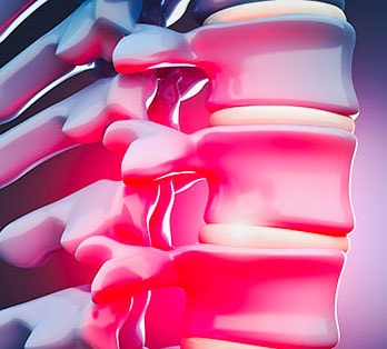
An intervertebral disc is a strong shock absorber that connects one vertebral bone to the next. The discs are the cushions between each vertebra of the spine. Each disc has a strong outer ring of outer fibers, called the annulus, and a soft, jelly-like center, called the nucleus pulposus. The annulus is the strongest area of the disc and connects each vertebra together. The strong annular fibers contain the nucleus and distribute pressure evenly across the disc. The nucleus, or center of the disc, is hydrated and serves as the main shock absorber. The nucleus absorbs the impact of the body's daily activities and keeps the two vertebrae separated.
Just like other ligaments, the discs can be injured and torn. The annulus can rupture anywhere around the disc and can leak the inner disc material which is very irritating to nerves and can cause intense pain and inflammation. The outer 1/3 of the disc’s annular ring is highly innervated with pain fibers. Thus, if a tear involves the outer 1/3 it may be extremely painful. This tear will heal with scar tissue over time but is more prone to future tears and injury.
Annular Tear Causes
Most annular tears are caused by the natural aging process but can be caused by trauma as well. Since the neck and back are responsible for bearing most of a person’s body weight, they are susceptible to a great deal of wear over time. By the age of 30, 60% of people have discs that show signs of degneration. By the age of 60, 100% of people have signes of disc degeneration including herniations and tears.
Annular Tear Symptoms
Predominant symptoms in many patients with an annular tear in the low back include midline back pain in excess of leg pain. Pain is often worse when sitting compared to standing, and positions that load pressure on the disc (coughing, sneezing, forward bending and lifting) tend to aggravate symptoms.
Diagnosing Annular Tears
After a physical assessment, a doctor will typically perform one or more diagnostic tests such as an MRI to localize the likely source of your pain. Occasionally your doctor may suggest a discography test which contrast is injected directly into the discs, to see if the disc is truly the source of pain. The procedure is performed under fluoroscopic x-ray guidance and allows your physician to assess the integrity of the disc and monitor your experience of pain at the same time. A CT scan is performed after the procedure to visualize the structure of the disc, and to evaluate for annular tears.